

Identify the Bony Posterior Wall of the Pelvis
Are you looking for a comprehensive guide on how to identify the bony posterior wall of the pelvis? Look no further! In this article, I’ll provide you with all the information you need to accurately locate and understand this crucial anatomical structure.
The bony posterior wall of the pelvis plays a significant role in providing support and stability to the pelvic region. It consists of several bones, including the sacrum and coccyx, which form the base of the spine. Understanding its location is crucial for various medical professionals, such as orthopedic surgeons, radiologists, and physical therapists.
To identify the bony posterior wall of the pelvis, it’s essential to have a clear understanding of its landmarks. These include palpating and visualizing specific points on the body, such as the sacral promontory and coccyx. Additionally, utilizing imaging techniques like X-rays or CT scans can provide detailed insight into this region.
In this comprehensive guide, I will walk you through step-by-step instructions on how to identify these landmarks accurately. Whether you’re a healthcare professional or simply curious about human anatomy, this article will equip you with valuable knowledge regarding the bony posterior wall of the pelvis. So let’s dive in and explore this fascinating topic together!
Understanding the structure and landmarks of this important anatomical feature is crucial for healthcare professionals, students, or anyone interested in pelvic anatomy.
The pelvis is a remarkable part of our body that plays a vital role in providing support and protection to our reproductive organs, urinary system, and digestive system. Its bony posterior wall forms an integral component of the pelvis, contributing to its overall stability and function.
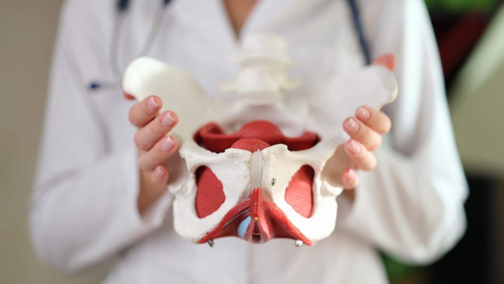
To identify the bony posterior wall accurately, we need to familiarize ourselves with key landmarks. The sacrum is one such landmark – a triangular bone located at the base of the spine between the two hip bones. It plays a significant role in connecting the spinal column with the pelvic girdle.
Another essential landmark is the coccyx, commonly known as our tailbone. Situated below the sacrum, it consists of several fused vertebrae that provide additional support to our pelvis.
Additionally, understanding how these structures relate to other parts of our body is crucial. The sacroiliac joint connects each side of the sacrum with its respective ilium bone – forming strong connections that stabilize our pelvis during weight-bearing activities.
By utilizing imaging techniques such as X-rays or CT scans, healthcare professionals can further enhance their ability to visualize and assess any potential abnormalities or injuries affecting this area. These diagnostic tools play an invaluable role in ensuring accurate identification and diagnosis.
In conclusion, identifying the bony posterior wall of the pelvis requires knowledge about essential landmarks like the sacrum and coccyx. Understanding their relationship within our skeletal framework allows healthcare professionals and enthusiasts alike to appreciate its significance in maintaining pelvic stability.